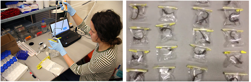
Approximately 96 hours prior to exposure, individuals are placed in whirlpac bags with DNA-free water for one hour to collect a mucosome sample. After the hour, the water is decanted into 15 ml conical tubes and frozen at -80 Celsius. Mucosome samples are sent to collaborator, Doug Woodhams, at the University of Massachussetts, Boston, for in vitro Bsal inactivation assays.
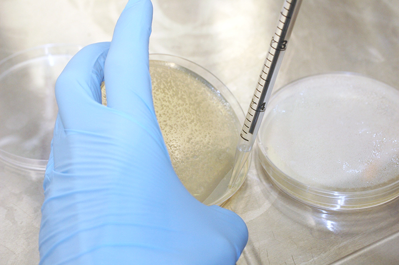
Biosafety Level 2 protocols are followed to ensure containment of Bsal within area dedicated for experimentation. Bsal cultures are provided by Laura Reinert and Louise Rollins-Smith at Vanderbilt University. The day of exposure, Bsal zoospores are suspended in deionized water using three separate washes: two using 3ml water, the final using 1 ml. The first and last wash are removed immediately after swirling; the second is left on the plate for 20 minutes before being pipetted off the plate.
After removing the washes from the Bsal culture plate, they are vacuum filtered using 20 micrometer cell microsieves.
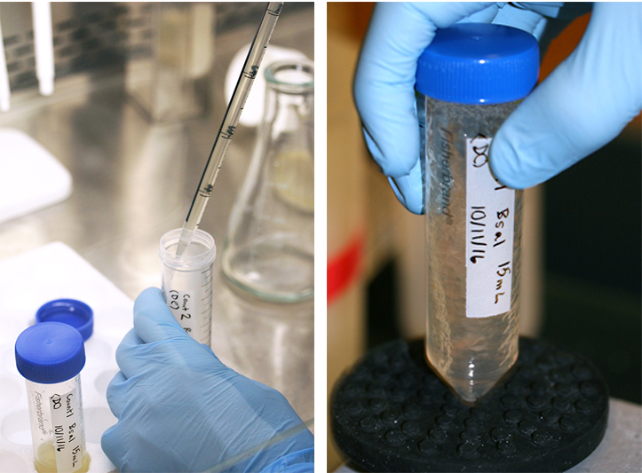
The resulting extact is transferred into 50ml falcon tubes and then vortexed to suspend any zoospores that have settled.
A small portion of the extract is then diluted 10x in microcentrifuge tubes for enumeration.
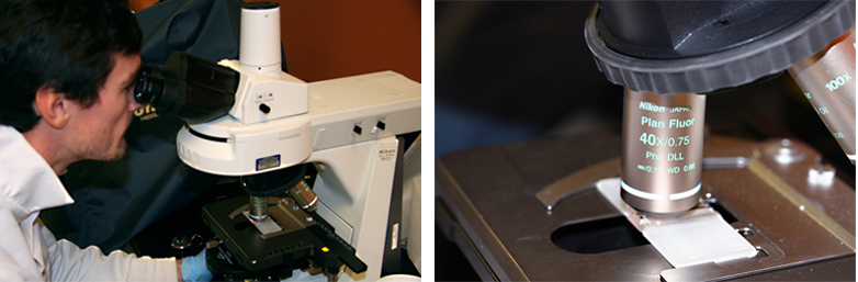
The diluted extract is again vortexed to mix the suspension, and then 10 microliters of the suspension is placed in a hemocytometer for counting zoospores.
The zoospores are counted in quadruplicate (i.e., from four cardinal locations on the slide) under 40x magnification to determine the average zoospores per ml of the filtered washes.
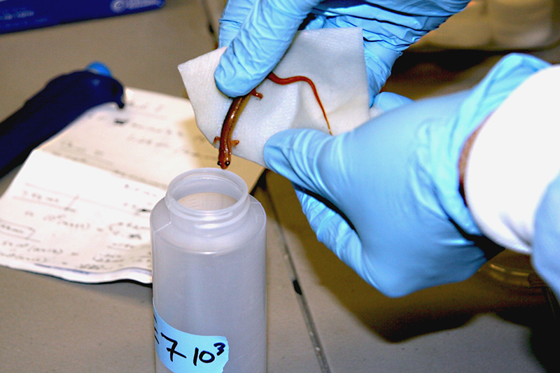
After enumeration, the wash extract is serially diluted into stock solutions of 5×10³-6 zoospores per ml.
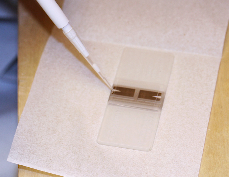
In order to inoculate the animals, 1ml of each stock solution is added to 9ml of water in a 100ml honey tube (the exposure container), resulting in doses of 5×10³-6 zoospores. (Honey tubes and honey tube lids were purchased at the Cary Company.)
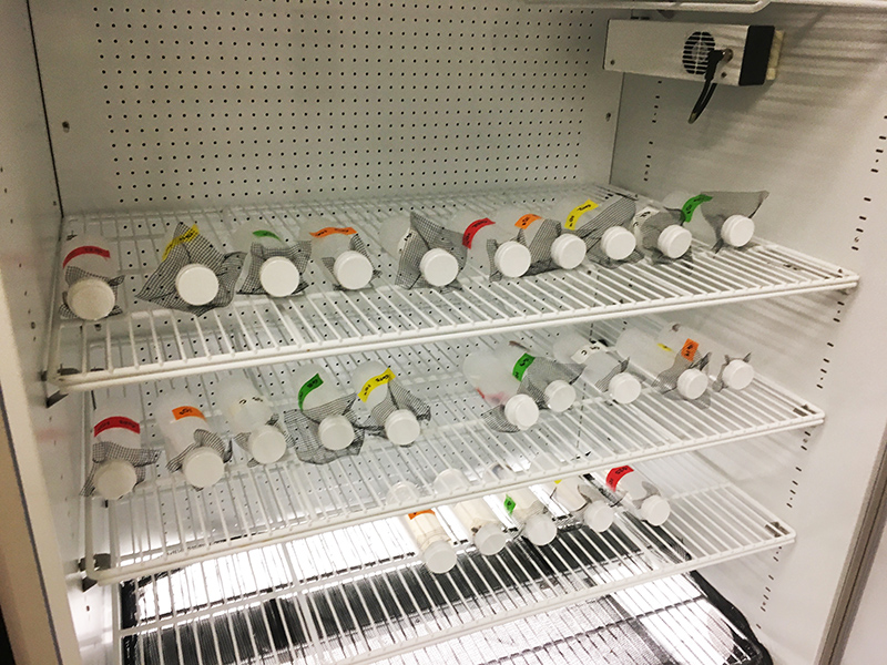
After the inoculum is prepared, individuals are gently transferred from housing containers into the exposure tubes. The tubes are covered with fine-meshed screen to prevent escape and then capped with the flip top open to allow airflow. The target sample size for an experiment is five-10 animals per dose for four zoospore doses, and an additional five-10 control animals.
The exposure containers are placed on their side in a randomized block design in an incubator set to 15 Celsius (the temperature at which Bsal grows best in cell culture). The animals are left in the exposure containers for 24 hours, during which time the honey tubes are rotated every 6 hours to ensure animals are in contact with the inoculum.
After 24 hours in the exposure containers, the inoculum is poured into a bucket with granulated chlorine and the animals are placed into food service containers (14L x 11W x 6H cm) with an unbleached paper towel moistened with dechlorinated water and a cover object. (Food service containers were purchased on Amazon.)
The housing containers are placed in a randomized block design in incubators at 15 Celsius and are checked twice daily for 42. Animals are fed equivalent to 2 percent of their body mass and have their housing containers cleaned every three days. During cleaning, containers are placed in a 15 g/L sodium hypochlorite solution for a minimum of 10 minutes to inactivate Bsal shed from the animal, then rinsed with dechlorinated water overnight.
For aquatic species, 1ml of Bsal stock solution is pipetted directly onto the back of the animal, and the animals are placed in isolation containers filled with 3L of dechlorinated water at 15 Celsius for 24 hours.
After 24 hours in the isolation containers, animals are placed into 38L aquaria filled with dechlorinated water at 15 Celsius in a flow-through system that cycles at 400ml/min and drains into a reservoir with clorine tabs for inactivating Bsal. As with terrestrial species, aquatic species are fed every three days and provided a PVC cover object.
Swab samples are collected starting four days post-exposure following the protocol for chytrid fungus swab sampling (video), then every six days thereafter until the end of the experiment. When swabbing, each of the hands, as well as the ventral surface are swabbed five times each, for a total of 30 swab passes per individual per sampling event. Additional swab samples are taken if lesioning is observed. All animal handling follows approved Institutional Animal Care and Use protocols.
After 42 days any surviving animals are humanely euthanized following the guidelines put forth by the American Veterinary Medical Association. At death (if during the test) or immediately prior to euthanasia, a final swab sample is collected from each animal. Two toe samples and a sample of ventral skin are collected from dead animals and frozen separately at -80 Celsius for qPCR analyses to test for Bsal, and animals are inspected for clinical signs of Bsal chytridiomycosis (e.g., skin sloughing, lesions, etc.).
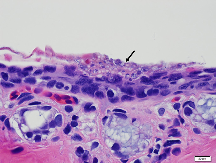
Bsal chytridiomycosis is then confirmed via histopathology. For example, this lesion from an infected animal contains multiple encysted Bsal thalli, as indicated by the black arrow.
Bsal infection is measured following the protocol of Blooi et al. (2013). Fungal DNA is extracted from frozen swab and tissue samples using Qiagen Dneasy Blood and Tissue Kits and analyzed via qPCR in duplicate with a positive and a negative control. A sample is considered positive if it has a threshold cycle (CT) value of 50 or lower. The number of DNA copies is determined using a standard curve generated using gBlock synthetic DNA.